
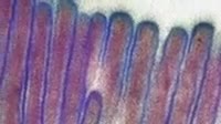
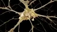
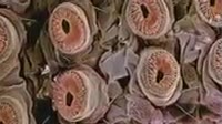
Retina rod cells. Coloured scanning electron micrograph SEM of a freeze_fractured section through a retina, revealing the structure of its photoreceptors. Photoreceptors with these rod_like outer parts green are named rod cells rather than cone cells and contain the protein rhodopsin visual purple that aids vision in dim light. The inner parts orange lead to the nuclei blue red, top. Empty spaces in this layer show Muller cells that provide a fibrous support for photoreceptors. Light triggers signals in the photoreceptors after passing through a neuron layer above nuclei, not seen. These signals travel to the neurons and on to the brain.

Details
WebID:
C00596575
Clip Type:
RM
Super High Res Size:
1920X1080
Duration:
000:22.000
Format:
QuickTime
Bit Rate:
25 fps
Available:
download
Comp:
200X112 (0.00 M)
Model Release:
NO
Property Release
NO
 Loading