
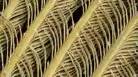
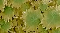
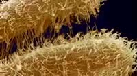
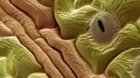
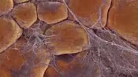
Microvilli in duodenum. Coloured transmission electron micrograph TEM of a section through the human duodenum, showing microvilli on the surface epithelium lining. The duodenum is the first part of the small intestine. The microvilli dark blue appear as tiny projections from the surface of the epithelial cells at lower frame. Microvilli are present on two specialised cell types that comprise the duodenal epithelium. One type, goblet cells, secrete mucus, a second type, secretory cells, secrete digestive enzymes and an alkaline fluid. The existence of microvilli serves to maximise the duodenum磗 surface area and hence its capacity to secrete.


Details
WebID:
C00596555
Clip Type:
RM
Super High Res Size:
1920X1080
Duration:
000:17.000
Format:
QuickTime
Bit Rate:
25 fps
Available:
download
Comp:
200X112 (0.00 M)
Model Release:
NO
Property Release
NO
 Loading