
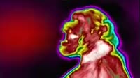
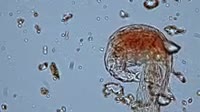
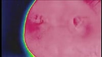
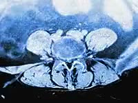
Meiosis cell division. Labelled animation showing the stages of meiotic cell division (meiosis). At the start of the clip, the chromosomal genetic material (red and blue) is shown in the nucleus (orange). Within the nucleus is the nucleolus (round). The nucleus is bounded by the nuclear membrane. Outside the nucleus, structures called centrioles (black, left and right) are shown. During meiotic cell division, chromosomes form matched pairs called homologous chromosomes, one from the father and one from the mother (paternal and maternal). During meiosis, genetic material is swapped between the maternal and paternal chromosomes, with the swapping shown here in close up. The view then zooms out to show the whole cell, where the nuclear membrane has dissolved and the chromosomes have lined up at the centre of the cell. The centrioles have formed a spindle that pulls the chromosomes apart as the cell divides. The nuclear membrane then reforms, ending what is known as Meiosis I. The result is two haploid cells with half the number of chromosomes compared to the original diploid cell. The next stage, known as Meiosis II, is where the two haploid cells divide normally, creating four haploid cells. These cells (gametes) and the process of meiosis itself, are a key part of sexual reproduction. The cell division stages are named prophase, metaphase, anaphase and telophase. For this animation without labels, see clip K004/6345.


Details
WebID:
C01843295
Clip Type:
RM
Super High Res Size:
1920X1080
Duration:
00:00:52.000
Format:
QuickTime
Bit Rate:
25 fps
Available:
download
Comp:
200X112 (0.00 M)
Model Release:
NO
Property Release
No
 Loading