
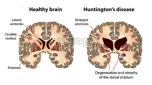
Illustration comparing coronal sections of a healthy brain and a brain in Huntington's disease. The brain in Huntington's disease shows enlarged anterior horns of the lateral ventricles, degeneration and atrophy of the dorsal striatum.
| px | px | dpi | = | cm | x | cm | = | MB |
Details
Creative#:
TPG32656691
Source:
達志影像
Authorization Type:
RF
Release Information:
須由TPG 完整授權
Model Release:
N/A
Property Release:
N/A
Right to Privacy:
No
Same folder images:
artworkbasalgangliabraincaudatenucleuscentralnervoussystemcerebralhemispherechromosomecorpusstriatumgenegeneticshealthhuntingtonschoreaillustrationlentiformnucleusmedicalmedicinemovementdisordernervoussystemneuronaldegenerationneuropathyputamencoronalsectionhuntingtonsdiseaseautosomaldominantchromosome4httgenehuntingtinproteinwhitebackgroundplainbackgroundnobodyno-one
4artworkautosomalbackgroundbackgroundbasalbraincaudatecentralcerebralchoreachromosomechromosomecoronalcorpusdegenerationdiseasedisorderdominantgangliagenegenegeneticshealthhemispherehtthuntingtinhuntingtonshuntingtonsillustrationlentiformmedicalmedicinemovementnervousnervousneuronalneuropathyno-onenobodynucleusnucleusplainproteinputamensectionstriatumsystemsystemwhite
 Loading