
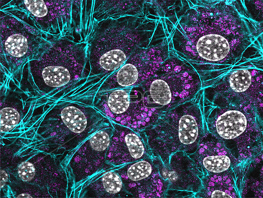
Immunofluorescence micrograph of murine fibroblasts stained with an actin cytoskeleton probe (teal), lysotracker probe (purple), and nuclear DAPI (white). Focal planes were achieved with an optical apotome.
| px | px | dpi | = | cm | x | cm | = | MB |
Details
Creative#:
TOP26739563
Source:
達志影像
Authorization Type:
RM
Release Information:
須由TPG 完整授權
Model Release:
N/A
Property Release:
N/A
Right to Privacy:
No
Same folder images:
 Loading