
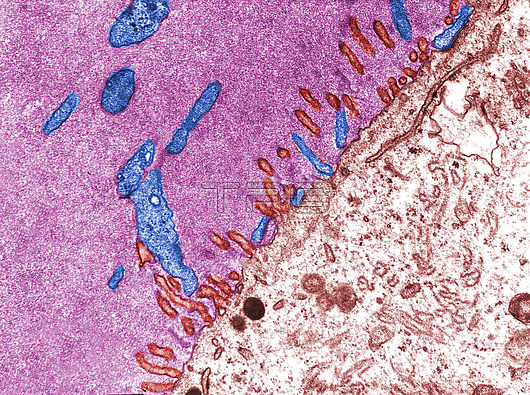
Coloured transmission electron micrograph (TEM) showing the zona pellucida (pink) and the oocyte cytoplasm (brown). Short microvilli of the oocyte (red) and longer and thicker microvilli (blue) of follicular cells (some reaching the oocyte surface) are crossing the zona pellucida.
| px | px | dpi | = | cm | x | cm | = | MB |
Details
Creative#:
TOP26739318
Source:
達志影像
Authorization Type:
RM
Release Information:
須由TPG 完整授權
Model Release:
N/A
Property Release:
N/A
Right to Privacy:
No
Same folder images:
biologicalbiologycellcellultrastructurecytologicalcytologyelectronmicroscopefalsecolourfalse-colouredfemalegranulosacellhistologicalhistologymicrographmicroscopemicroscopyoocyteovaryreproductivesystemtemtransmissionelectronmicrographmicroscopynobodyno-onetransmissionelectronmicroscopeultrastructurezonapellucida
biologicalbiologycellcellcellcolourcytologicalcytologyelectronelectronelectronfalsefalse-colouredfemalegranulosahistologicalhistologymicrographmicrographmicroscopemicroscopemicroscopemicroscopymicroscopyno-onenobodyoocyteovarypellucidareproductivesystemtemtransmissiontransmissionultrastructureultrastructurezona
 Loading