
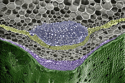
Scanning electron micrograph of a corner, 2mm wide, of the square stem of red dead nettle, Lamium purpureum. The picture shows several cell types; each has its role to play. New cells arise by mitosis (cell division) in the layer of cambium (yellow). Some merely expand in size, to produce parenchyma (grey); a space-filling tissue with no specialised purpose. The blue region is a vascular bundle, containing pipes that convey water up from the roots (xylem), or nutrient sap down from the leaves (phloem). The purple layer of small elongated cells with thick walls is the collenchyma. Its siting and structure provides strength to the stem. The green outer layer is the epidermis; a waterproof skin. The epidermis of leaves contains stomatal pores that allow the plant to breathe (not seen here). All the tissues of higher plants develop by the process illustrated here; cell differentiation.
| px | px | dpi | = | cm | x | cm | = | MB |
Details
Creative#:
TOP26709386
Source:
達志影像
Authorization Type:
RM
Release Information:
須由TPG 完整授權
Model Release:
N/A
Property Release:
N/A
Right to Privacy:
No
Same folder images:
 Loading