
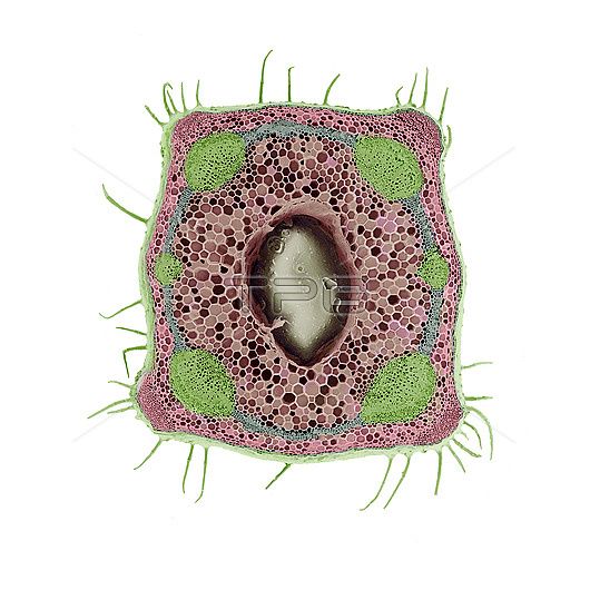
Scanning electron micrograph of a stem of red dead nettle, Lamium purpureum in cross section, illustrating different tissue types. The picture shows the square outline of a hollow stem, with hairs on its surface. The coloured ring comprises six (green) water-conducting vascular bundles linked by a layer of cambium (blue). To the outside of the ring is the cortex; to the inside is the pith (both pink). The pith consists of parenchyma - cells with no specialised function, that may contain chloroplasts or starch grains. During growth, the pith cells degenerate, giving rise to the hollow stem here. The hollow space is not empty; it is filled with water and remnants of cell walls. The cortex is also parenchymatous, but at the extreme corners, it has differentiated to form a tissue of small cells called collenchyma. This is a structural specialisation giving mechanical support to the stem
| px | px | dpi | = | cm | x | cm | = | MB |
Details
Creative#:
TOP26709378
Source:
達志影像
Authorization Type:
RM
Release Information:
須由TPG 完整授權
Model Release:
N/A
Property Release:
N/A
Right to Privacy:
No
Same folder images:
 Loading