
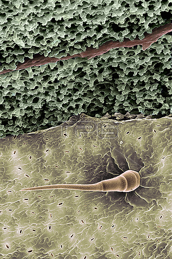
Scanning electron micrograph of the underside of a garden Zinnia leaf. The picture shows the lower surface of the leaf and its inner cell structure, seen from below. The surface of the epidermis shows one glandular hair (brown), 0.7mm long, and many small pores (stomata). Stomata allow air to enter the leaf, and control the evaporation of water from its interior. Here, they are open. If a plant is water stressed, they close, and the leaf wilts. In the upper part of the picture, the epidermis has been removed. This reveals internal cells (green) with many air spaces - the spongy mesophyll. The picture shows a vein (red/brown) passing through the mesophyll; this carries water from the roots. Spongy mesophyll is a photosynthetic tissue, but the main photosynthesis is performed by rows of palisade cells exposed to light under the upper epidermis of the leaf - "behind" the green cells here.
| px | px | dpi | = | cm | x | cm | = | MB |
Details
Creative#:
TOP26624800
Source:
達志影像
Authorization Type:
RM
Release Information:
須由TPG 完整授權
Model Release:
N/A
Property Release:
N/A
Right to Privacy:
No
Same folder images:
 Loading