
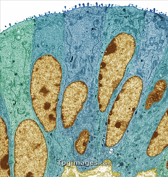
Fallopian tube, coloured transmission electron micrograph (TEM). Section through non-ciliated columnar epithelium from a fallopian tube. The ciliated cells are not shown here. Large prominent nuclei are present (brown) with microvilli visible at the cell surface (top). Magnification: x1100 when printed at 10 centimetres wide.
| px | px | dpi | = | cm | x | cm | = | MB |
Details
Creative#:
TPG05326995
Source:
達志影像
Authorization Type:
RF
Release Information:
須由TPG 完整授權
Model Release:
NO
Property Release:
NO
Right to Privacy:
No
Same folder images:
oviductfallopiantubehumancellnucleusmicrovillusfallopiantubeoviductanatomybiologyhistologyreproductioncytologycellbilogyfemalecolouredtemtransmissionelectronmicroscopeanatomicalbiologicalcellscolumnarepitheliaepithelialepitheliumfalse-colourfalse-colouredhealthyhistologicalmicrovillinon-ciliatednormalnucleireproductivesecretorysectionsectionedtracttransmissionelectronmicrograph"
"anatomicalanatomybilogybiologicalbiologycellcellcellscolouredcolumnarcytologyelectronelectronepitheliaepithelialepitheliumfallopianfallopianfalse-colourfalse-colouredfemalehealthyhistologicalhistologyhumanmicrographmicroscopemicrovillimicrovillusnon-ciliatednormalnucleinucleusoviductoviductreproductionreproductivesecretorysectionsectionedtemtracttransmissiontransmissiontubetube
 Loading