
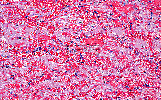
Light micrograph of red blood cells and fibrin. The red blood cells (many small bright red dots) clump together with fibrin (light pink) in a blood clot. Scattered fibroblast nuclei (blue elongated dots) are also seen, indicating organisation or the cellular reaction to a clot that has been present in the body for some time. Haematoxylin and eosin stained tissue section. Magnification: 200x when printed at 10cm.
| px | px | dpi | = | cm | x | cm | = | MB |
Details
Creative#:
TOP29676259
Source:
達志影像
Authorization Type:
RM
Release Information:
須由TPG 完整授權
Model Release:
N/A
Property Release:
N/A
Right to Privacy:
No
Same folder images:
pathologypathologicalmedicinemedicaldiseasedisorderconditionabnormalunhealthyanatomicpathologybloodcirculatorysystembloodclotcoagulationcardiovascularthrombusredbloodcellsfibrinorganizingbloodclothistopathologicalhistopathologymicroscopylmlightmicrographslidehematoxylinandeosinhaematoxylinandeosinhumanbodyanatomytissuecellsnobodyno-one
abnormalanatomicanatomyandandbloodbloodbloodbloodbodycardiovascularcellscellscirculatoryclotclotcoagulationconditiondiseasedisordereosineosinfibrinhaematoxylinhematoxylinhistopathologicalhistopathologyhumanlightlmmedicalmedicinemicrographmicroscopyno-onenobodyorganizingpathologicalpathologypathologyredslidesystemthrombustissueunhealthy
 Loading