
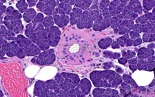
Light micrograph of cells of the parotid gland. The acinar cells (present in most areas of the image) contain many dark purple granules. A duct (pink circle) is present in the centre of the image. A few fat cells (white circles) are also present, as well as a blood vessel containing red blood cells (lower left corner). Haematoxylin and eosin stained tissue section. Magnification: 200x when printed at 10cm.
| px | px | dpi | = | cm | x | cm | = | MB |
Details
Creative#:
TOP29676227
Source:
達志影像
Authorization Type:
RM
Release Information:
須由TPG 完整授權
Model Release:
N/A
Property Release:
N/A
Right to Privacy:
No
Same folder images:
anatomyanatomicalparotidglandsalivaryglandacinarcellsductmajorsalivaryglanddigestivesystemexocrineexocrineglanddigestionheadandneckheadandneckENTotolaryngologybiologybiologicalhistologyhistologicalmicroscopylmlightmicrographslidehematoxylinandeosinhaematoxylinandeosinhumanbodyanatomytissuecellsnormalhealthynobodyno-one
ENTacinaranatomicalanatomyanatomyandandandandbiologicalbiologybodycellscellsdigestiondigestiveducteosineosinexocrineexocrineglandglandglandglandhaematoxylinheadheadhealthyhematoxylinhistologicalhistologyhumanlightlmmajormicrographmicroscopyneckneckno-onenobodynormalotolaryngologyparotidsalivarysalivaryslidesystemtissue
 Loading