
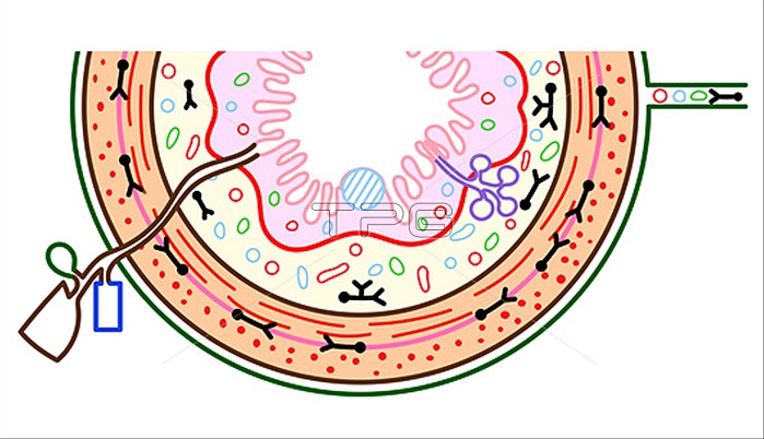
Gut, illustration. General organisation of the gastrointestinal tract. The innermost mucosa (pink) includes the muscularis mucosae (red line). Next is the submucosa (yellow) followed by layers of smooth muscle (orange) of the muscularis externa. Most of the gut is surrounded by mesentery (dark green). Arteries (red), veins (blue), lymphatics (green) and nerves (black) are shown. Lymphoid follicles (hatched blue) of the ileum, and submucosal Brunner?™s glands (purple) of the duodenum are also shown. Ducts from the liver, gall bladder and pancreas empty into the duodenum.
| px | px | dpi | = | cm | x | cm | = | MB |
Details
Creative#:
TOP29389355
Source:
達志影像
Authorization Type:
RM
Release Information:
須由TPG 完整授權
Model Release:
Not Available
Property Release:
Not Available
Right to Privacy:
No
Same folder images:
 Loading