
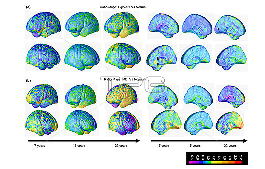
Coloured magnetic resonance imaging (MRI) scan mapping of grey matter maturation across the cortex in subjects between the ages of 7 and 22 years. The brains at top left are from before and after the subjects were diagnosed with paediatric bipolar disorder. The brains at bottom left are from subjects diagnosed with atypical psychosis (multi-dimensionally impaired, MDI) who did not go on to develop bipolar disorder. The brains at right are from healthy subjects. The bipolar brains show increased grey matter (yellow, orange and red) over the left temporal cortex and decreased grey matter (blue) in the anterior cingulate cortex. The changes were more striking after illness onset. The pattern was similar in MDI subjects and so may reflect dysregulation in general.
| px | px | dpi | = | cm | x | cm | = | MB |
Details
Creative#:
TOP29217729
Source:
達志影像
Authorization Type:
RM
Release Information:
須由TPG 完整授權
Model Release:
Not Available
Property Release:
Not Available
Right to Privacy:
No
Same folder images:
 Loading