
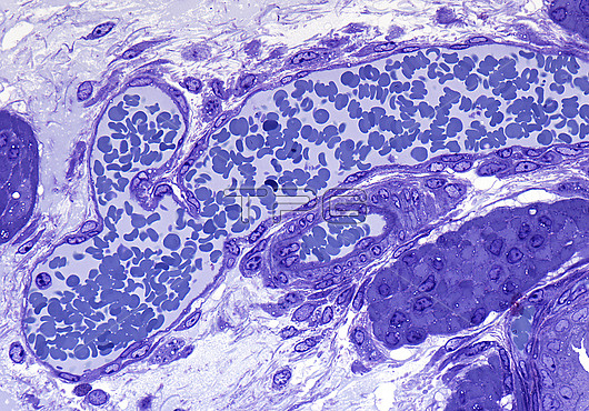
Venule, light micrograph. Longitudinal section through a venule with a smaller joining tributary vessel. Venules have a thin wall of inner endothelium external to which are fibroblasts and mesenchymal cells collectively referred to as pericytes. An adjacent arteriole of smaller calibre shows a thicker wall comprised chiefly of smooth muscle. Epoxy resin section, Toluidine blue stain. Magnification: x350 when width printed at 10cm.
| px | px | dpi | = | cm | x | cm | = | MB |
Details
Creative#:
TOP28610430
Source:
達志影像
Authorization Type:
RM
Release Information:
須由TPG 完整授權
Model Release:
n/a
Property Release:
n/a
Right to Privacy:
No
Same folder images:
 Loading