
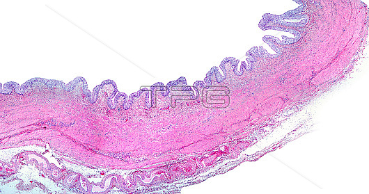
Light micrograph of a urinary bladder wall showing a very folded mucosa layer, lined by transitional epithelium (urothelium) resting on a connective tissue lamina propria. A muscular layer with smooth muscle cells arranged in thin bundles separated by connective tissue septa, and a very thin adventitia layer with large blood vessels can be seen.
| px | px | dpi | = | cm | x | cm | = | MB |
Details
Creative#:
TOP28014694
Source:
達志影像
Authorization Type:
RM
Release Information:
須由TPG 完整授權
Model Release:
N/A
Property Release:
N/A
Right to Privacy:
No
Same folder images:
 Loading