
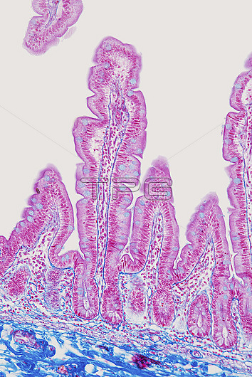
Light micrograph of a section through the mucosa of the jejunum, the middle part of the small intestine, which runs from the stomach to the large intestine. It is where digestion begins and nutrients are absorbed into the blood. The interior (lumen) is lined with villi, which are folds in the intestinal surface that greatly increase the surface area for absorption. Within the villi are goblet cells, which secrete mucus (blue). Also within the surface are crypts of Lieberkuhn (rings of cells), which secrete enzymes into the lumen that help to digest the food. Across bottom is connective tissue (blue) containing blood vessels (red). Magnification: x200 when printed at 15cm tall.
| px | px | dpi | = | cm | x | cm | = | MB |
Details
Creative#:
TOP27148062
Source:
達志影像
Authorization Type:
RM
Release Information:
須由TPG 完整授權
Model Release:
N/A
Property Release:
N/A
Right to Privacy:
No
Same folder images:
 Loading