
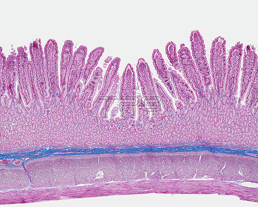
Light micrograph of a section through a duodenum, the beginning of the small intestine, which runs from the stomach to the large intestine. It is where digestion begins and nutrients are absorbed into the blood. The interior (lumen, top) is lined with villi, which are folds in the intestinal surface that greatly increase the surface area for absorption. Also within the surface are crypts of Lieberkuhn (red-lined ovoids), which secrete enzymes into the lumen that help to digest the food. Underlying this layer is the muscularis mucosae (thin violet line) and connective tissue (blue). Across bottom are layers of smooth muscle and then circular and longitudinal muscles that contract and relax to move food through the duodenum. Magnification: x40 when printed at 15 centimetres wide.
| px | px | dpi | = | cm | x | cm | = | MB |
Details
Creative#:
TOP27147866
Source:
達志影像
Authorization Type:
RM
Release Information:
須由TPG 完整授權
Model Release:
N/A
Property Release:
N/A
Right to Privacy:
No
Same folder images:
 Loading