
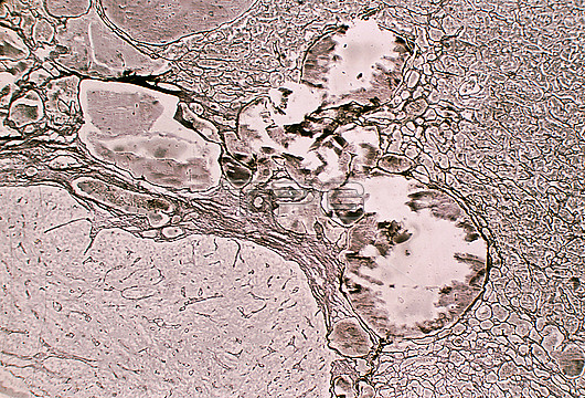
Reticular fibres in human pituitary gland, light micrograph. This is the border between the anterior lobe (right), intermediate lobe (centre) and posterior lobe (left) of the pituitary. It has been stained with the silver method to highlight reticular fibres. With this method, the differences in the organization pattern of each of these lobes stand out. The anterior lobe shows a cell cords pattern. The intermediate lobe contains large vesicles and cysts (whose content appears fragmented by cutting artifacts). The posterior lobe has poor reticulin development, limited to the wall of the blood vessels.
| px | px | dpi | = | cm | x | cm | = | MB |
Details
Creative#:
TOP26812123
Source:
達志影像
Authorization Type:
RM
Release Information:
須由TPG 完整授權
Model Release:
N/A
Property Release:
N/A
Right to Privacy:
No
Same folder images:
 Loading