
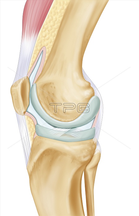
Representation of the knee joint on an internal lateral view of the right leg. At the center of the illustration are the two articular surfaces in gray / light green, the articular surface of the distal epiphysis of the femur and the internal meniscus. placed on the tibial head. The articular capsule is filled with synovium represented in pale blue with the cruciate ligaments in transparency, surrounded by its pink membrane. The ligaments of the knee are distinguished in light gray. The patella is found in front of the knee. the articulation on the left of the drawing and the head of the fibula behind the right of the drawing ... The patella is attached up to the lower tendon of the quadriceps muscle and down to the tibia with the patellar ligament.
| px | px | dpi | = | cm | x | cm | = | MB |
Details
Creative#:
TOP25709751
Source:
達志影像
Authorization Type:
RM
Release Information:
須由TPG 完整授權
Model Release:
N/A
Property Release:
N/A
Right to Privacy:
No
Same folder images:
 Loading