
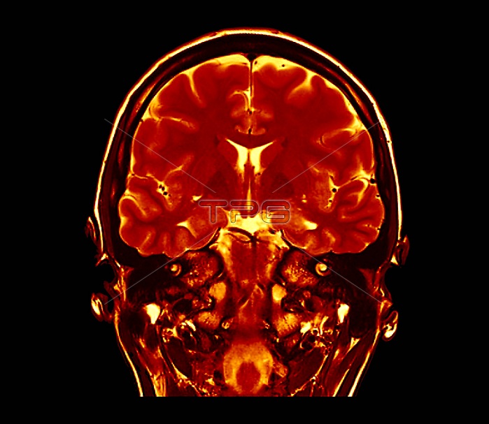
Human brain. Coloured coronal magnetic resonance imaging (MRI) scan through the brain of a 30-year-old man. This is a T2-weighted scan. The scan has passed vertically through the head, as seen from the front, at the level of the ears. The scan shows the normal brain structures, including parts of the lateral ventricles (white, centre) and the folds of the cerebrum. Below the brain, on either side of the head, are the structures of the inner ear.
| px | px | dpi | = | cm | x | cm | = | MB |
Details
Creative#:
TOP25477131
Source:
達志影像
Authorization Type:
RM
Release Information:
須由TPG 完整授權
Model Release:
N/A
Property Release:
N/A
Right to Privacy:
No
Same folder images:
ANATOMICALBIOLOGICALBLACKBACKGROUNDCORONALCUTOUTCUTOUTSCUT-OUTCUT-OUTSCUTOUTCUTOUTSEARSFALSE-COLOUREDHEALTHYINNEREARLATERALVENTRICLESMAGNETICRESONANCEIMAGINGNO-ONENOBODYNORMALSECTIONSECTIONEDSPIN-SPINSPINALT2RELAXATIONT2SCANT2WEIGHTEDTHIRTIESVENTRICLESORGANBRAINVENTRICLESPINEEARSKULLBONEHUMANBODYHEADBIOLOGYANATOMYADULT30S30MALEMANMRISCANCOLOUREDSCANNER
30MALE30SADULTANATOMICALANATOMYBACKGROUNDBIOLOGICALBIOLOGYBLACKBODYBONEHUMANBRAINCOLOUREDCORONALCUTCUTCUT-OUTCUT-OUTSCUTOUTCUTOUTSEAREAREARSFALSE-COLOUREDHEADHEALTHYIMAGINGINNERLATERALMAGNETICMANMRINO-ONENOBODYNORMALORGANOUTOUTSRELAXATIONRESONANCESCANSCANSCANNERSECTIONSECTIONEDSKULLSPIN-SPINSPINALSPINET2T2T2THIRTIESVENTRICLEVENTRICLESVENTRICLESWEIGHTED
 Loading