
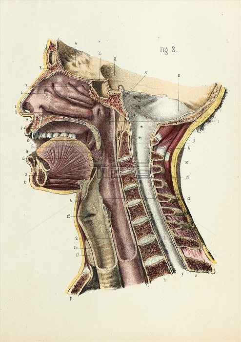
Mouth and neck anatomy, 1866 illustration. This midline dissection passes vertically through the head and neck, showing structures such as the nose, tongue, backbone, spinal cord, oesophagus and trachea. This is figure 2 from plate 4 from the third volume of 'Atlas d'anatomie descriptive du corps humain' (1844-1866) by French anatomists Constantin Bonamy and Paul Broca. This work described the anatomy of the human body with over 250 hand-coloured lithographs. The illustrations were by Emile Beau, with the text by Bonamy and Broca. The three volumes were bound as four books in 1866 when the atlas was completed. This page is from the third book 'Digestion', a republication of the section on the digestive system that was first published in 1850.
| px | px | dpi | = | cm | x | cm | = | MB |
Details
Creative#:
TOP24040471
Source:
達志影像
Authorization Type:
RM
Release Information:
須由TPG 完整授權
Model Release:
No
Property Release:
No
Right to Privacy:
No
Same folder images:
 Loading