
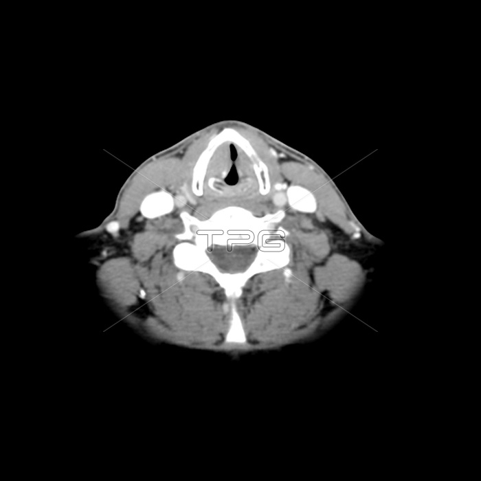
This axial (cross sectional) contrast enhanced CT shows what a paralyzed left (on your right) vocal cord would look like whilst holding breath. This person has a mass in the left side of the mediastinum of the chest. The paralyzed left (on your right) vocal cord is unable to move medially whilst holding breath. The normal right (on your left) vocal cord moves medially, as one would expect with a normally mobile vocal cord. Also note the more medial position of the right (on your left) arytenoid cartilage.
| px | px | dpi | = | cm | x | cm | = | MB |
Details
Creative#:
TOP22287472
Source:
達志影像
Authorization Type:
RM
Release Information:
須由TPG 完整授權
Model Release:
N/A
Property Release:
No
Right to Privacy:
No
Same folder images:
 Loading