
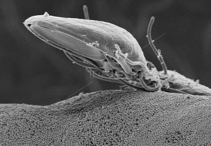
Scanning electron micrograph (SEM) depicted a Giardia specie intestinal protozoan on the microvillous border of intestinal epithelial cells. Each small circular profile under the protozoan represents the rounded tip of a single microvillous, and it is estimated that 2000 to 3000 microvilli cover the surface of a single intestinal epithelial cell. The ventral adhesive disk, which facilitates adherence to the intestinal surface, can be seen on the underside of the organism. The protozoan Giardia causes the diarrheal disease called giardiasis. Giardia species exist as free-swimming (by means of flagella) trophozoites, and as egg-shaped cysts. It is the cystic stage, which facilitates the survival of these organisms under harsh environmental conditions. The cyst is considered the infective form, and disease is often transmitted by drinking contaminated water. As depicted in these SEMs, in the intestine, cysts are stimulated to liberate trophozoites. Cysts can be shed in fecal material, and can, thereafter, remain viable for several months in appropriate environmental conditions. Cysts can also be transferred directly from person-to-person, as a result of poor hygiene.
| px | px | dpi | = | cm | x | cm | = | MB |
Details
Creative#:
TOP22237443
Source:
達志影像
Authorization Type:
RM
Release Information:
須由TPG 完整授權
Model Release:
N/A
Property Release:
No
Right to Privacy:
No
Same folder images:
 Loading