
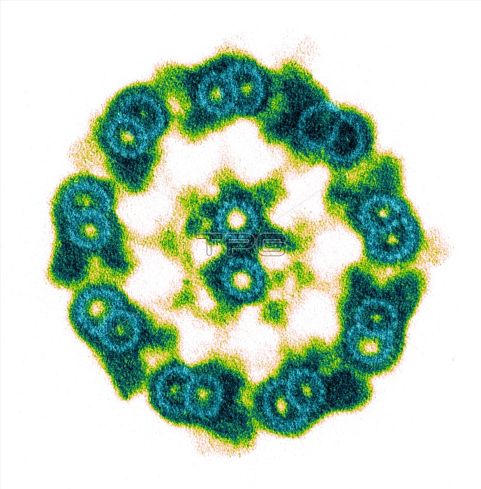
Color enhanced transmission electron micrograph of the cilia of Tetrahymena thermophila are used not only for feeding and propelling the cell through the medium but also for sensing the environment around them. This image is of an isolated axoneme with the membrane removed and stained with tannic acid. The tannic acid allows the visualization of the individual tubulin heterodimers that make up the walls of the microtubule doublets. The central pair and A tubules of the microtubule doublets are made up of 13 tubulin heterodimers whereas the B tubules of the microtubule doublets are made up of 11 heterodimers. Magnification 125,000X.
| px | px | dpi | = | cm | x | cm | = | MB |
Details
Creative#:
TOP22231859
Source:
達志影像
Authorization Type:
RM
Release Information:
須由TPG 完整授權
Model Release:
N/A
Property Release:
No
Right to Privacy:
No
Same folder images:
 Loading