
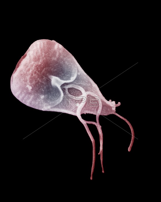
This scanning electron micrograph (SEM) reveals some of the external ultrastructural details displayed by a flagellated Giardia lamblia protozoan parasite. G. lamblia is the organism responsible for causing the diarrheal disease giardiasis. Once an animal or person has been infected with Giardia intestinalis, the parasite lives in the intestine and is passed in the stool. Because the parasite is protected by an outer shell, it can survive outside the body and in the environment for long periods of time. Cysts are resistant forms and are responsible for transmission of giardiasis. Both cysts and trophozoites can be found in the feces (diagnostic stages). The cysts are hardy and can survive several months in cold water. Infection occurs by the ingestion of cysts in contaminated water, food, or by the fecal-oral route (hands or fomites). In the small intestine, excystation releases trophozoites (each cyst produces two trophozoites). Trophozoites multiply by longitudinal binary fission, remaining in the lumen of the proximal small bowel where they can be free or attached to the mucosa by a ventral sucking disk. Encystation occurs as the parasites transit toward the colon. The cyst is the stage found most commonly in non-diarrheal feces. Because the cysts are infectious when passed in the stool or shortly afterward, person-to-person transmission is possible. While animals are infected with Giardia, their importance as a reservoir is unclear.
| px | px | dpi | = | cm | x | cm | = | MB |
Details
Creative#:
TOP22225237
Source:
達志影像
Authorization Type:
RM
Release Information:
須由TPG 完整授權
Model Release:
N/A
Property Release:
No
Right to Privacy:
No
Same folder images:
 Loading