
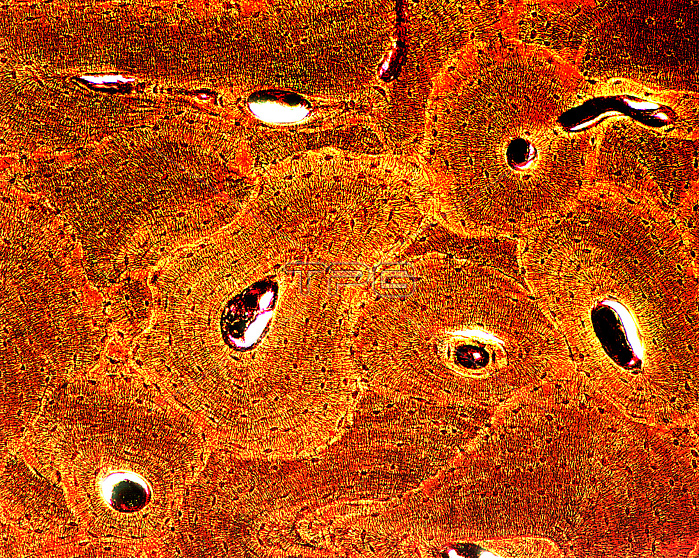
Light micrograph showing a section through compact bone of a mature long bone at low magnification. The micrograph shows a number of Haversian systems seen in cross section. The center of a Haversian system contains a Haversian canal through which blood vessels and nerves traverse. Each Haversian system consists of concentric lamellae of bone tissue along with osteocytes between lamellae. The osteocytes appear as densely stained, elongate cell bodies. Present between Haversian systems are interstitial lamellae that represent the remnants of pre-existing Haversian systems. Schmorl's stain. Magnification: x175.
| px | px | dpi | = | cm | x | cm | = | MB |
Details
Creative#:
TOP22223197
Source:
達志影像
Authorization Type:
RM
Release Information:
須由TPG 完整授權
Model Release:
N/A
Property Release:
No
Right to Privacy:
No
Same folder images:
 Loading