
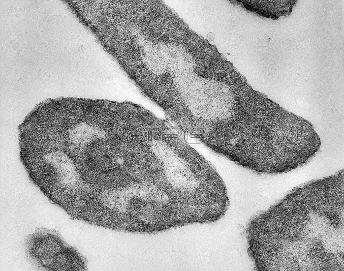
TEM autoradiograph of mating bacteria. The Hfr (high frequency recombination) cell, is the large cell at the top of picture. It was labeled with the radioctive tracer H3-Thymine. The F- cell (at bottom) was unlabeled, and mating took place in cold medium. The autoradiographic grains indicate the presence of lableled DNA in the nucleus of the Hfr, and its transfer to the nucleus of the F- cell. Unlike a normal F+ cell, hfr strains will, upon conjugation with a F- cell, attempt to transfer their entire DNA through the mating bridge, not to be confused with the pilus. This occurs because the F factor has integrated itself via an insertion point in the bacterial chromosome. Due to the F factor's inherent nature to transfer itself during conjugation, the rest of the bacterial genome is dragged along with it, thus making such cells very useful and interesting in terms of studying gene linkage and recombination. Because the genome's rate of transfer through the pilus is constant, molecular biologists and geneticists can use Hfr strain of bacteria to study genetic linkage and map the chromosome. The procedure commonly used for this is called interrupted mating. Mag. 140,000x.
| px | px | dpi | = | cm | x | cm | = | MB |
Details
Creative#:
TOP22219866
Source:
達志影像
Authorization Type:
RM
Release Information:
須由TPG 完整授權
Model Release:
No
Property Release:
No
Right to Privacy:
No
Same folder images:
 Loading