
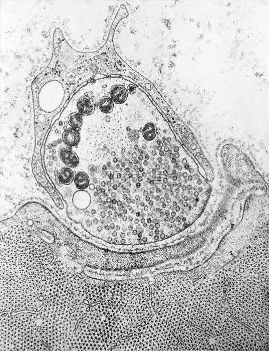
Transmission electron micrograph of a frog myoneural junction prepared by freeze substitution. The nerve terminal is covered by a portion of Schwann cell (above), and is separated from the muscle fiber (below) by a synaptic cleft containing a conspicuous external lamina. The nerve ending's cytoplasm contains a few microtubules, several mitochondria, a large aggregation of synaptic vesicles, and a number of neurofilaments.
| px | px | dpi | = | cm | x | cm | = | MB |
Details
Creative#:
TOP22219556
Source:
達志影像
Authorization Type:
RM
Release Information:
須由TPG 完整授權
Model Release:
N/A
Property Release:
No
Right to Privacy:
No
Same folder images:
 Loading