
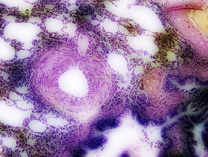
Artery in lung tissue. Digitally enhanced light micrograph of a thin section through lung tissue with haematoxylin and eosin. This view shows an artery (centre left) surrounded by alveoli. At lower right is a bronchiole, lined with ciliated columnar epithelial cells. At top right is cartilage supporting a bronchus, one of the major airways of the lung, keeping it from collapsing. Alveoli are the tiny air sacs of the lungs where gases are exchanged between the air and the blood in the blood vessels.
| px | px | dpi | = | cm | x | cm | = | MB |
Details
Creative#:
TOP19654064
Source:
達志影像
Authorization Type:
RM
Release Information:
須由TPG 完整授權
Model Release:
N/A
Property Release:
N/A
Right to Privacy:
No
Same folder images:
alveolibronchiolebrightfieldbronchicartilagecilliatedcolumnarepitheliumepitheliealepitheliumhyalinecartilagemicroscopyno-onenobodyphotomicrographrespirationthinsectioneosinhaematoxylinhematoxylinbiologicalanatomicalhistologicalstainstainedsectionsectionedarterialvascularcirculationcirculatorysystemcellsbronchialnormalhealthycellularTISSUECELLARTERYBLOODVESSELALVEOLUSBRONCHIOLEHUMANBODYLUNGRESPIRATORYSYSTEMBIOLOGYANATOMYHISTOLOGYLIGHTMICROGRAPHLMLIGHTMICROSCOPE
ALVEOLUSANATOMYARTERYBIOLOGYBLOODBODYBRONCHIOLEHUMANCELLHISTOLOGYLIGHTLIGHTLMLUNGMICROGRAPHMICROSCOPERESPIRATORYSYSTEMTISSUEVESSELalveolianatomicalarterialbiologicalbrightfieldbronchibronchialbronchiolecartilagecartilagecellscellularcilliatedcirculationcirculatorycolumnareosinepitheliealepitheliumepitheliumhaematoxylinhealthyhematoxylinhistologicalhyalinemicroscopyno-onenobodynormalphotomicrographrespirationsectionsectionsectionedstainstainedsystemthinvascular
 Loading