
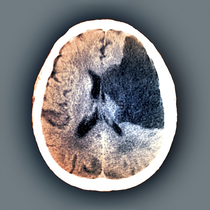
Brain in stroke, CT scan. Coloured computed tomography (CT) scan of an axial section through the brain of a 62-year-old person affected by hemiplegia (paralysis) of the right side of the body following a stroke (cerebral vascular accident, cva). Here, a large hypodense area (dark, upper right) can be seen affecting the sylvian and ventricular regions. Strokes occur when the blood supply to an area of the brain is restricted, or cut off completely, resulting in cell death of the affected tissue.
| px | px | dpi | = | cm | x | cm | = | MB |
Details
Creative#:
TOP17481090
Source:
達志影像
Authorization Type:
RM
Release Information:
須由TPG 完整授權
Model Release:
N/A
Property Release:
N/A
Right to Privacy:
No
Same folder images:
6262-year-old62-years-oldabnormalbraincerebralvascularaccidentcoloredcolouredcomputedtomographyconditionctcutoutcutoutscut-outcut-outscutoutcutoutscvadiagnosticimagingdisorderfalse-colouredhealthcarehemiplegiamedicalmedicineneurologicalneurologyno-onenobodyradiographyradiologicalradiologyrightscansectionsectionedsixtiesstrokesylvianunhealthyventricleventriclesventricularx-rayxrayxrays
6262-year-old62-years-oldabnormalaccidentbraincerebralcoloredcolouredcomputedconditionctcutcutcut-outcut-outscutoutcutoutscvadiagnosticdisorderfalse-colouredhealthcarehemiplegiaimagingmedicalmedicineneurologicalneurologyno-onenobodyoutoutsradiographyradiologicalradiologyrightscansectionsectionedsixtiesstrokesylviantomographyunhealthyvascularventricleventriclesventricularx-rayxrayxrays
 Loading