
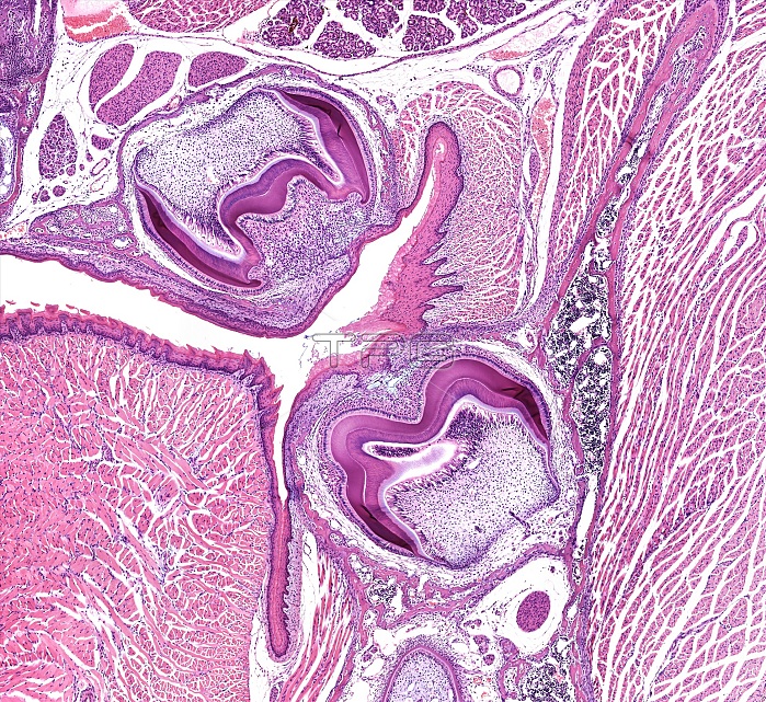
Light microscopy of fetal teeth in an advanced stage of development. Each tooth is located below the gumline in a socket formed in the bone of the jaw. Part of the tongue is to the left. The growing crown (magenta curvatures) of the teeth is well defined, and show a sculptured appearance suggestive of the structure of molar teeth. The outermost layer is the growing enamel next to which is the dentin. The core of each tooth will become the pulp or root cavity where nerves and blood vessels supply the tooth. The region between the tooth and the jawbone will form the periodontal ligament that anchors a tooth in the socket-type recess. Magnification x40 when printed at 10 cm.
| px | px | dpi | = | cm | x | cm | = | MB |
Details
Creative#:
TOP14988181
Source:
達志影像
Authorization Type:
RM
Release Information:
須由TPG 完整授權
Model Release:
N/A
Property Release:
No
Right to Privacy:
No
Same folder images:
 Loading