
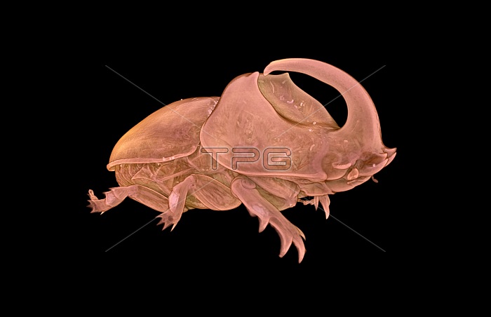
Rhinoceros beetle (Oryctes boas), micro-CT scan. The micro-CT scanner was used to investigate the internal anatomy of this large beetle. This image was acquired using X-ray micro-Computed Tomography (also known as micro-CT). A micro-CT scanner projects a beam of X-rays through the sample onto a detector panel. Images are collected over a 36MB0?rotation and these are then reconstructed to form a virtual 3D model of the specimen. This virtual object that can be viewed from all angles and sliced open or digitally dissected. Image by Dan Sykes.
| px | px | dpi | = | cm | x | cm | = | MB |
Details
Creative#:
TOP13789479
Source:
達志影像
Authorization Type:
RM
Release Information:
須由TPG 完整授權
Model Release:
N/A
Property Release:
No
Right to Privacy:
No
Same folder images:
 Loading