
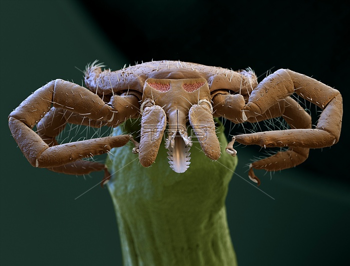
Sheep tick. Coloured scanning electron micrograph (SEM) of a female sheep tick (Ixodes ricinus) showing its mouthparts. The mouthparts of a tick consist of three visible components. The two outer jointed parts are highly mobile palps. Between the palps, at centre, is a rod-shaped structure, the hypostome. The palps do not enter the skin of the host while the tick is feeding, it is the hypostome that is inserted into the host's skin. The backward-pointing projections on the hypostome prevent easy removal of the tick. Magnification: x35 when printed at 10 centimetres across.
| px | px | dpi | = | cm | x | cm | = | MB |
Details
Creative#:
TOP12392501
Source:
達志影像
Authorization Type:
RM
Release Information:
須由TPG 完整授權
Model Release:
No
Property Release:
No
Right to Privacy:
No
Same folder images:
IXODESRICINUSSHEEPTICKANIMALARACHNIDLEAFBIOLOGYZOOLOGYCOLOUREDSEMSCANNINGELECTRONMICROSCOPEANATOMICALANATOMYARACHNOLOGYBIOLOGICALBLOODSUCKINGBLOODSUCKERCASTORBEANTICKFALSE-COLOUREDFAUNAFEEDINGFEMALEHYPOSTOMEINVERTEBRATEIXODOIDEAMOUTHPARTMOUTHPARTSNATUREPALPPALPSSCANNINGELECTRONMICROGRAPHTICKTICKMOUTHPARTSUNDERNEATHUNDERSIDEWILDLIFEZOOLOGICAL
ANATOMICALANATOMYANIMALARACHNIDARACHNOLOGYBEANBIOLOGICALBIOLOGYBLOODBLOODSUCKERCASTORCOLOUREDELECTRONELECTRONFALSE-COLOUREDFAUNAFEEDINGFEMALEHYPOSTOMEINVERTEBRATEIXODESIXODOIDEALEAFMICROGRAPHMICROSCOPEMOUTHPARTMOUTHPARTSMOUTHPARTSNATUREPALPPALPSRICINUSSCANNINGSCANNINGSEMSHEEPSUCKINGTICKTICKTICKTICKUNDERNEATHUNDERSIDEWILDLIFEZOOLOGICALZOOLOGY
 Loading