
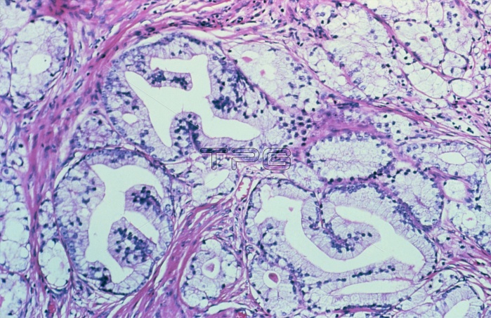
Light micrograph showing benign prostatic hyperplasia. Hyperplasia describes an increase in cell numbers due to cell division. In the prostate gland, the inner portion & its surrounding fibromuscular stroma may become hyperplastic. Here, the acini (white spaces) are lined with tall, purple-stained cells with small basal nuclei; the glandular lining appears buckled into papillary folds. Adjacent acini are separated by thickened fibromuscular connective tissue (red stain). Since the inner (paraurethral) part of the gland is involved, compression of the urethra is common. This accounts for the symptoms of urinary retention that are common in elderly men.
| px | px | dpi | = | cm | x | cm | = | MB |
Details
Creative#:
TOP11722225
Source:
達志影像
Authorization Type:
RM
Release Information:
須由TPG 完整授權
Model Release:
NO
Property Release:
NO
Right to Privacy:
No
Same folder images:
 Loading