
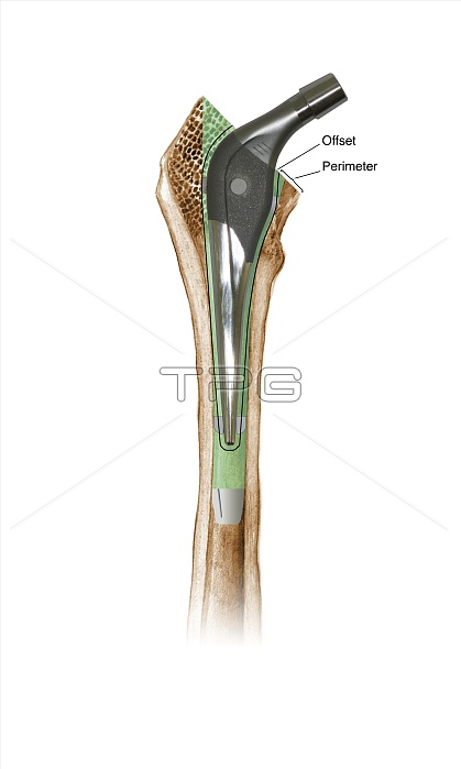
Prosthetic hip joint. Cutaway diagram of a femur (thigh bone) showing a femoral component of a hip prosthesis. This component is implanted in the femur after the head of the femur has been surgically removed. The other components of the hip joint are a rounded ball to fit into the socket implanted in the patient's pelvis (not shown). This allows the patient to regain mobility, and is usually done to treat severe osteoarthritis or a broken hip. This is an SHP prosthesis, using bone cement (green). The 'offset' and 'perimeter' parameters are labelled. For this diagram with Gruen zones, see C016/6776.
| px | px | dpi | = | cm | x | cm | = | MB |
Details
Creative#:
TOP11716550
Source:
達志影像
Authorization Type:
RM
Release Information:
須由TPG 完整授權
Model Release:
NO
Property Release:
NO
Right to Privacy:
No
Same folder images:
 Loading