
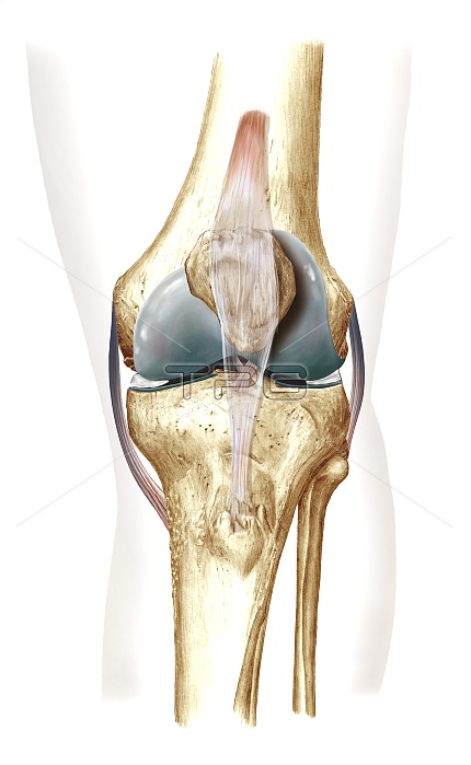
Knee bones and ligaments. Artwork of a frontal view of the knee joint, showing bones, ligaments, tendons and cartilage. The bones are the femur (thigh bone, top), patella (knee-cap, upper centre), and the lower leg bones, the tibia (lower centre) and the fibula (lower right). The quadriceps femoris muscle tendon passes over the patella, becoming the patellar ligament that attaches to the tibia. The medial collateral ligament (left) joins femur and tibia. The fibular collateral ligament (right) joins femur and fibula. Two short ligaments (centre) join femur and tibia: the posterior and anterior cruciate ligaments. The menisci (grey) are cartilage pads cushioning the joint.
| px | px | dpi | = | cm | x | cm | = | MB |
Details
Creative#:
TOP11249970
Source:
達志影像
Authorization Type:
RM
Release Information:
須由TPG 完整授權
Model Release:
No
Property Release:
No
Right to Privacy:
No
Same folder images:
 Loading