
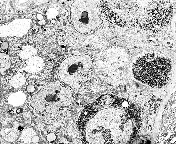
Testicular cancer. Transmission electron micrograph (TEM) of a section through a testis (testicle), showing neoplastic germ cells (dark, circular), the precursors of testicular cancer. The cells are characteristically round and commonly found next to the basal lamina of the seminiferous tubule's epithelium. Above these premalignant cells are three Sertoli cell nuclei but other germ cells are absent. Also termed carcinoma-in-situ (CIS), the abnormal germ cells often have large amounts of glycogen (black dots) in the cytoplasm, as seen here. Magnification x5600, when printed 10 centimetres wide.
| px | px | dpi | = | cm | x | cm | = | MB |
Details
Creative#:
TOP10393044
Source:
達志影像
Authorization Type:
RM
Release Information:
須由TPG 完整授權
Model Release:
No
Property Release:
No
Right to Privacy:
No
Same folder images:
 Loading