
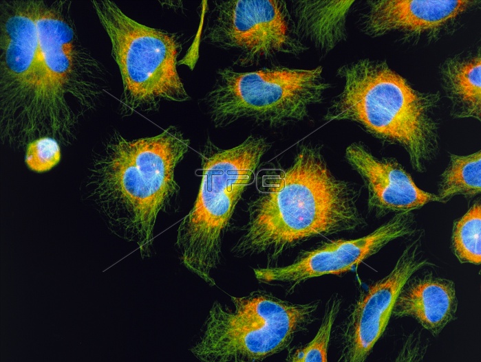
Mitosis. Immunofluorescent light micrograph of HeLa cancer cells undergoing mitotic division. Many of these cells are in the resting stage between cell division, known as Interphase. This is when the spindle structures, which play a vital role in cell division, are formed. The formation of spindles is seen in several cells as two light spots around the blue-stained nucleus. At top left corner, a cell in the final stage of mitosis, telophase, is seen. This is when the nucleus divides into two identical daughter nuclei. Immunofluorescence uses antibodies to attach fluorescent dyes to specific molecules in cells. Magnification: x125 at 6x4.5cm size.
| px | px | dpi | = | cm | x | cm | = | MB |
Details
Creative#:
TOP10222023
Source:
達志影像
Authorization Type:
RM
Release Information:
須由TPG 完整授權
Model Release:
N/A
Property Release:
N/A
Right to Privacy:
No
Same folder images:
 Loading