
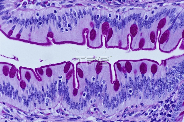
Intestinal villi. Light micrograph of the surfaces of two adjacent villi on the lining of the small intestine. Each fold of the wall of the small intestine is covered with these small projections, which serve to increase the surface area for the absorption of nutrients from the gut lumen. Villi are mainly composed of columnar epithelial cells (purple) interspersed with goblet cells (pink). The goblet cells secrete mucous, which lines the surface of the membrane. The regularly ordered nuclei (blue/purple) of the columnar cells are easily visible. Magnification: x340 when printed 10cm wide.
| px | px | dpi | = | cm | x | cm | = | MB |
Details
Creative#:
TOP10220647
Source:
達志影像
Authorization Type:
RM
Release Information:
須由TPG 完整授權
Model Release:
N/A
Property Release:
N/A
Right to Privacy:
No
Same folder images:
 Loading