
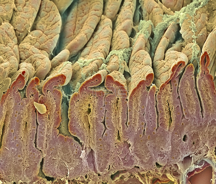
Intestinal lining. Coloured scanning electron micrograph (SEM) of a freeze-fractured intestinal surface. The fracture plane (lower frame) has cut down through the surface, revealing the deep folds called villi. The intestinal surface (orange) is where food is digested. It consists of a layer of surface (epithelial) cells (red), supported by connective tissue (orange). This connective tissue is seen in the fracture plane at the core of each fold (villus). The folds increase the area for the absorption of food. This tissue is from a part of the small intestine called the duodenum. Magnification unknown.
| px | px | dpi | = | cm | x | cm | = | MB |
Details
Creative#:
TOP10220629
Source:
達志影像
Authorization Type:
RM
Release Information:
須由TPG 完整授權
Model Release:
N/A
Property Release:
N/A
Right to Privacy:
No
Same folder images:
 Loading