
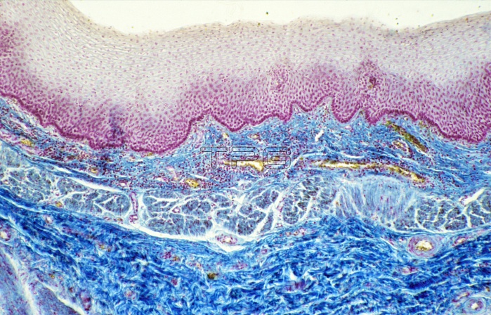
Light micrograph of a vertical section through the wall of a human oesophagus. Its lumen is lined by a thick stratified squamous epithelium (grey and magenta). This is supported by a thin layer of connective tissue known as the lamina propria. At bottom the muscularis layer is visible. It consists of smooth muscle and is divided into two layers. The inner one (centre) is formed by circular fibres and the outer one (bottom, dark blue) by longitudinal fibres. The action of these two muscle layers, opposed at right angles to one another, is the basis of the peristaltic contraction. Magnification: x25 at 35mm size.
| px | px | dpi | = | cm | x | cm | = | MB |
Details
Creative#:
TOP10220497
Source:
達志影像
Authorization Type:
RM
Release Information:
須由TPG 完整授權
Model Release:
N/A
Property Release:
N/A
Right to Privacy:
No
Same folder images:
 Loading