
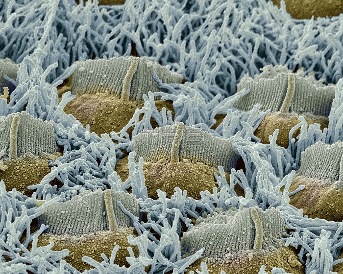
Foetal inner ear hair cells. Coloured scanning electron micrograph (SEM) of hair cells in the organ of Corti of a foetus. This delicate structure in the cochlea of the inner ear converts sound vibrations into nerve impulses. The three rows of outer hair cells have V-shaped groups of stereocilia, each with a central kinocilium. In the foetus they are surrounded by numerous microvilli (blue), which are resorbed in the adult. The hair cells are surrounded by endolymph fluid. As sound enters the ear it causes waves to form in the endolymph, which in turn cause these hairs to move. The movement is converted into nerve impulses that are passed to the brain.
| px | px | dpi | = | cm | x | cm | = | MB |
Details
Creative#:
TOP10220211
Source:
達志影像
Authorization Type:
RM
Release Information:
須由TPG 完整授權
Model Release:
N/A
Property Release:
N/A
Right to Privacy:
No
Same folder images:
 Loading