
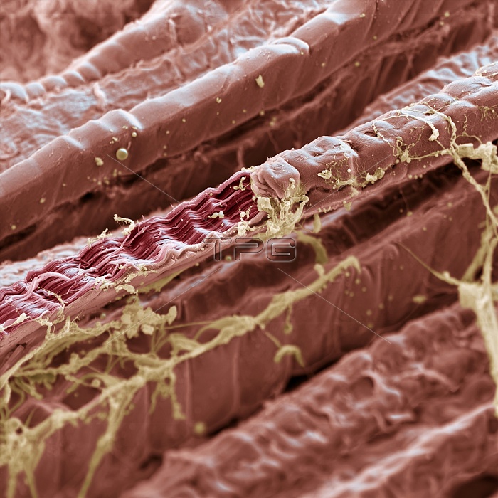
Skeletal muscle. Coloured scanning electron micrograph (SEM) of skeletal (striated) muscle. The muscle fibres are extremely long, cylindrical cells. One of the fibres has been torn open to expose thin threads called myofibrils. Myofibrils are formed of alternating bands of the proteins actin and myosin, which give the fibres a striped appearance. The proteins slide over each other to allow the muscle to contract. Connective tissue (yellow) forms a sheath known as the endomysium, which holds the muscle fibres together. Skeletal muscle attaches to the skeleton and is controlled by the voluntary nervous system. Magnification: x360 when printed 10 centimetres wide.
| px | px | dpi | = | cm | x | cm | = | MB |
Details
Creative#:
TOP10217542
Source:
達志影像
Authorization Type:
RM
Release Information:
須由TPG 完整授權
Model Release:
N/A
Property Release:
N/A
Right to Privacy:
No
Same folder images:
 Loading