
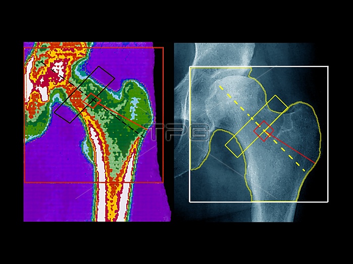
Hip scan and X-ray. Coloured bone densitometry scan (left) and an X-ray (right), of the same area of a left hip. The rectangle shows the neck of the femur (thigh bone). This connects the round head of the femur to its shaft. The head fits into its socket in the pelvis (upper left in each image). A bone densitometry scan uses X-rays to measure bone density. The bone density is colour-coded, ranging from blue/green (least dense) through to white (most dense). The average bone density of this menopausal patient is normal. This kind of scan is often used to detect decreased bone density (osteoporosis).
| px | px | dpi | = | cm | x | cm | = | MB |
Details
Creative#:
TOP10217202
Source:
達志影像
Authorization Type:
RM
Release Information:
須由TPG 完整授權
Model Release:
N/A
Property Release:
N/A
Right to Privacy:
No
Same folder images:
 Loading