
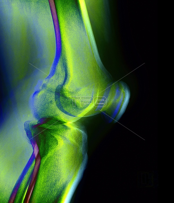
Knee joint. Coloured X-ray of a healthy human knee joint seen from the side. The bones are blurred to depict movement. The large femur (thigh-bone, top) articulates with the tibia (shin-bone, bottom) at the knee to form the knee joint. Next to the tibia is the smaller fibula bone (bottom left). The patella or kneecap (blurred, centre right) is a protective bone at the front of the knee held in position by muscles and tendons. Two discs of protective cartilage (not seen) cover the surfaces of the femur and tibia to reduce friction between these bones. This joint, the largest in the body, allows a backward-forward hinge movement with slight rotation.
| px | px | dpi | = | cm | x | cm | = | MB |
Details
Creative#:
TOP10217141
Source:
達志影像
Authorization Type:
RM
Release Information:
須由TPG 完整授權
Model Release:
N/A
Property Release:
N/A
Right to Privacy:
No
Same folder images:
 Loading