
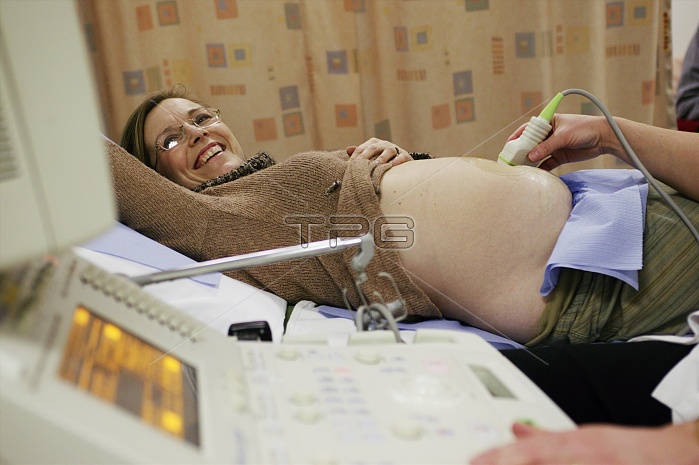
MODEL RELEASED. Pregnancy ultrasound. Pregnant woman having her swollen abdomen scanned by an ultrasound transducer (in hand). The transducer emits high-frequency sound waves and detects the reflected echoes. The results are used to build an image (which the mother is watching) of the unborn child in the womb. This safe, non-invasive method is used routinely in pregnancy to assess the growth and health of the developing foetus, and to detect any abnormalities. This is a standard anomaly scan, carried out when the mother is 20 weeks pregnant. The detection of a serious anomaly at this stage (halfway through pregnancy), allows surgery or a termination to be considered.
| px | px | dpi | = | cm | x | cm | = | MB |
Details
Creative#:
TOP10205076
Source:
達志影像
Authorization Type:
RM
Release Information:
須由TPG 完整授權
Model Release:
YES
Property Release:
N/A
Right to Privacy:
No
Same folder images:
 Loading