
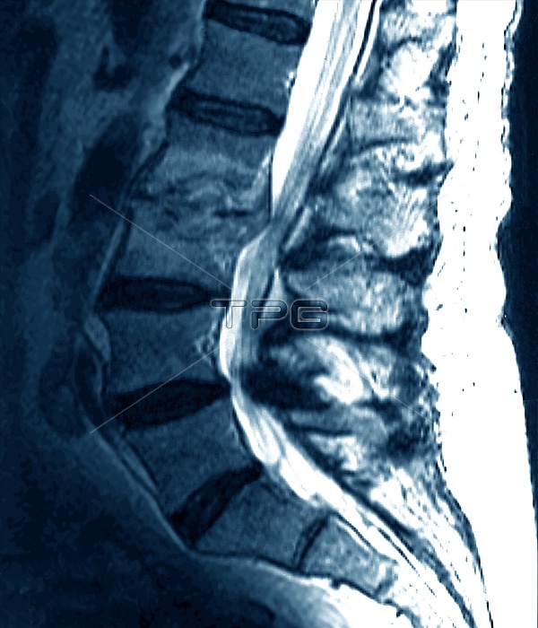
Spondylitis. Magnetic resonance imaging (MRI) scan of a sagittal (side) section through the lower spine of a 63-year-old patient suffering from spondylitis. The front of the body is at left. Intervertebral discs (black) separate the lumbar vertebrae (bones, blue). The space between vertebrae L2 and L3 (upper left) has narrowed, due to inflammation of the joint (spondylitis). Part of the joint is compressing the spinal cord (white, top centre to bottom right). Spondylitis causes pain and restriction of movement. Treatment includes physical therapy and anti- inflammatory drugs. In severe cases, surgery can relieve pressure on the spinal canal.
| px | px | dpi | = | cm | x | cm | = | MB |
Details
Creative#:
TOP10201363
Source:
達志影像
Authorization Type:
RM
Release Information:
須由TPG 完整授權
Model Release:
N/A
Property Release:
N/A
Right to Privacy:
No
Same folder images:
 Loading