
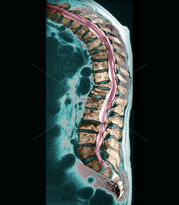
Osteoporosis. Coloured sagittal (side) magnetic resonance imaging (MRI) scan through the back of a 60-year-old patient with osteoporosis. The front of the body is at left. The spinal blocks of bone (vertebrae, brown) are seen enclosing the spinal cord (pink). Several vertebrae have collapsed due to the effects of osteoporosis. Good examples are just above centre and at upper centre. The effect of these collapses is a hunched spinal curvature (upper left). Osteoporosis is a decrease in bone density due to loss of bone material. It is mostly seen in the elderly and menopausal women. Surgical intervention can stabilise the spine. MRI scanning uses magnets and radio waves to image the body.
| px | px | dpi | = | cm | x | cm | = | MB |
Details
Creative#:
TOP10200298
Source:
達志影像
Authorization Type:
RM
Release Information:
須由TPG 完整授權
Model Release:
N/A
Property Release:
N/A
Right to Privacy:
No
Same folder images:
 Loading