
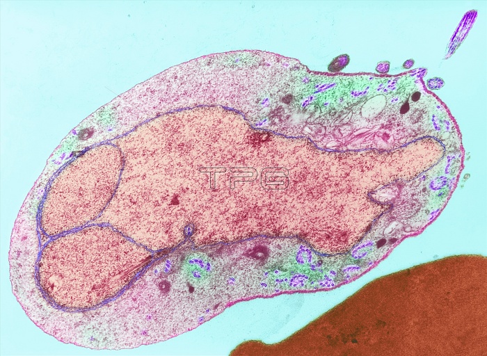
Malaria parasite. Image 4 of 10. Coloured transmission electron micrograph (TEM) of a sexual male malaria (Plasmodium sp.) microgametocyte in a mosquito (Anopheles sp.) gut. The microgametocyte releases male microgametes (purple, one at top right) after it is ingested by a mosquito feeding on an infected human. Each motile gamete possesses a flagellum built from fibrils (purple, paired) assembled near the gametocyte membrane. The male gametes fertilise the female macrogametocytes (not seen). Asexual reproduction then produces the stage that infects humans. Magnification: x5700 at 6x7cm size. For the malaria life cycle, see images M210/197-206.
| px | px | dpi | = | cm | x | cm | = | MB |
Details
Creative#:
TOP10199933
Source:
達志影像
Authorization Type:
RM
Release Information:
須由TPG 完整授權
Model Release:
N/A
Property Release:
N/A
Right to Privacy:
No
Same folder images:
 Loading